Podcast: Play in new window | Download
Subscribe: Apple Podcasts | Spotify | Android | Pandora | iHeartRadio | TuneIn | RSS

We discuss the role of point-of-care ultrasound in evaluating the patient with kidney injury and assessing volume status, with Abhilash Koratala (@nephroP), nephrologist, Director of Clinical Imaging for Nephrology at the Medical College of Wisconsin, and champion of nephrology-focused ultrasound.
Takeaway lessons
- A quick kidney and bladder ultrasound to rule out urinary obstruction is appropriate for most significant AKIs, maybe even if it was done previously (as obstruction can develop at any time).
- Ultrasound of the lungs and IVC help establish the presence of elevated filling pressures; if present, the VEXUS scan can be performed to establish the presence of venous congestion that might be contributing to kidney injury.
- Pulmonary edema as evidenced by B-lines establishes that the patient is not fluid tolerant, and suggests that further volume loading may be harmful. It increases the chance that AKI is due to congestive nephropathy as well, although each can also occur in isolation (and of course AKI can be a cause, leading to volume retention and then pulmonary edema).
- Abhilash does an 8-zone lung exam (2 anterior and 2 lateral zones on each side), which is plenty for cardiogenic pulmonary edema. He does not really count B-lines; if he sees B-lines in more than one dependent zone, he takes it as evidence the patient could be decongested.
- IVC is a reasonable method of estimating RAP; it is not reliable to gauge fluid responsiveness or other questions. The internal jugular vein is a good fallback if the IVC is untenable or seems unreliable, such as if bandages limit access, or the presence of cirrhosis (which alters local vasculature in unpredictable ways). Look for the highest point of distention and measure roughly from the sternal angle, adding it to the right atrial depth to approximate the CVP (usually ~5 cm although this is not very reliable).
- A non-plethoric IVC and absence of B-lines suggests a fluid tolerant patient. He uses the ACE guidelines of IVC >2.1 and <50% collapse with deep inspiration (sniff) to equate RAP ~15 mmHg.
- In the presence of elevated RAP, VEXUS helps determine whether that change is likely to be affecting organ perfusion by altering flow characteristics. Higher VEXUS scores are well-associated with risk of AKI.
- High RAP with a low VEXUS suggests that congestive nephropathy is not actively worsening renal function, whereas a higher VEXUS suggests the opposite. Serial VEXUS scans help track the progress of decongestion to dial in a patient to an optimal fluid balance.
- VEXUS is a right-sided heart parameter, so the state of the left heart’s filling may differ somewhat (e.g. as evidenced by lung markers like pulmonary edema—so track your B-lines too!). It is probably more precise and reliable than other markers like peripheral edema.
- Right and left heart filling should generally be well-linked. Venous congestion and elevated RAP usually indicate a well-filled LA as well, unless the lungs are acting as a significant resistor. If major PH is present, consider introducing measures like pulmonary vasodilators instead of further fluid loading; overdistending the RV will not help the LV.
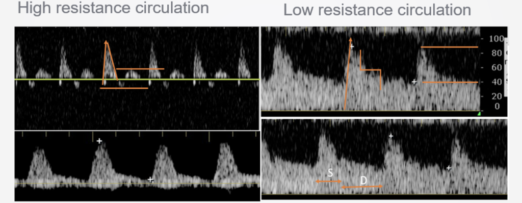
- Although portal vein pulsatility can usually move towards normal after optimal decongestion, hepatic vein waveforms may remain abnormal in some patients with TR, PH, etc. Flow chanegs in intrarenal vessels often lag behind other vessels, as renal edema takes time to resolve.
- Hepatic vein waveform may be permanently blunted in cirrhotics, confounding it somewhat. Distinguishing the systolic and diastolic waves can also be hard to identify without ECG synching; ECG is highly recommended when available.
- Portal vein is also probably unreliable in cirrhosis, due to portal hypertension and AVMs; it may be permanently pulsatile in some (although loss of pulsatility is still associated with decongestion). Since many cirrhotics have recurrent hospitalizations, you can compare against prior scans.
- Intrarenal vessels are technically difficult, especially when patients cannot perform a breath hold; critically ill patients have a failure rate here >20%. It is easier in stable patients. However, CKD patients may have abnormal waveforms at baseline.
- Overall, there should not be any disease state that falsely confounds ALL of the VEXUS vessels; while states like mechanical ventilation can increase flow changes and point to congestion, this is real congestion, not an artifact.
- Invasive monitoring like a CVP or PA catheter replaces some of the function of the VEXUS scan, but VEXUS helps determine the degree of organ impact at the numbers reported by these devices. High filling pressures generally are associated with congestion and low pressures are associated with its absence, but VEXUS is often helpful in the gray area. Non-invasive measurements of filling using echo, such as RVSP or E/A and E/E’ may have a supplemental role as well; the latter may help distinguish cardiogenic from non-cardiogenic pulmonary edema, but do not tell much of a story about systemic congestion.
- Femoral vein doppler could be a supplement to VEXUS, mainly when you cannot get or cannot trust one of the other vessels. It is farther from the heart, so may be less sensitive to changes in RAP. A normal femoral vein (continuous or mildly pulsatile) should probably not rule out venous congestion, but an abnormal femoral vein is very suggestive of it. This can sometimes be noted on routine DVT scans.
- Abhilash runs a cardiorenal clinic where he finds outpatient VEXUS very useful to establish and monitor volume status. It is more difficult to use in a dialysis clinic due to lack of privacy and high patient volumes; quick lung ultrasound (8 or even 4 zone) might be more useful here.
References
Episode 4 with Philippe Rola on the VEXUS scan
POCUS in AKI: Transcending boundaries: Unleashing the potential of multi-organ point-of-care ultrasound in acute kidney injury. Batool A, Chaudhry S, Koratala A. World J Nephrol 2023; 12(4): 93-103 [PMID: 37766842 DOI: 10.5527/wjn.v12.i4.93]
VEXUS for nephrologists: Koratala A, Reisinger N. Venous Excess Doppler Ultrasound for the Nephrologist: Pearls and Pitfalls. Kidney Med. 2022 May 19;4(7):100482. doi: 10.1016/j.xkme.2022.100482. PMID: 35707749; PMCID: PMC9190062.