Podcast: Play in new window | Download
Subscribe: Apple Podcasts | Spotify | Android | Pandora | iHeartRadio | TuneIn | RSS
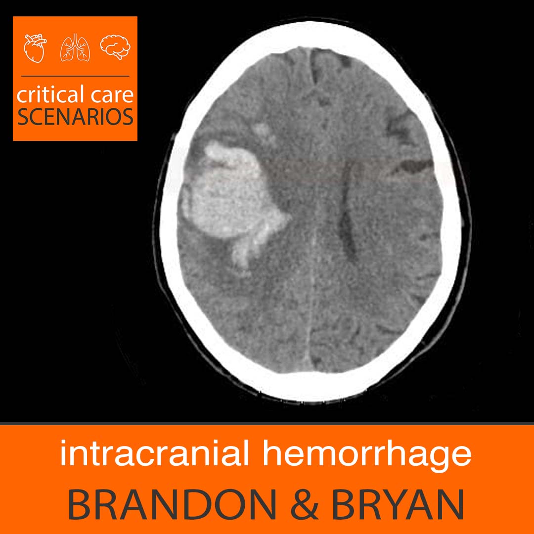
We review the basic care and workflow of caring for bread-and-butter intraparenchymal hemorrhage.
Learn more at the Intensive Care Academy!
Takeaway pearls
- IPH is technically a (hemorrhagic) stroke, which may mean it requires certain quality measures applicable to stroke certifications. (This doesn’t apply to traumatic bleeds, although it can sometimes be difficult to prove which ones are traumatic.)
- “ICH” could mean any intracranial hemorrhage, which includes intraparenchymal bleeds but also subdurals, epidurals, IVH, etc. Or it could mean “intracerebral,” which is synonymous with intraparenchymal. It’s a vague acronym.
- Which ICHs require ICU admission is a bit of a judgment call, mostly based on clinical picture and risk of expansion (size, anticoagulation status, etc). Some centers tend to admit nearly all of them, unless they are provably old and stable. Some may be quickly downgradable (or stay in the ED) if an initial stability scan is stable. Sometimes practical matters prevail, such as need for a continuous antihypertensive drip.
- The ICH score has some utility for prognostication and may be particularly helpful for determining disposition.
- ICU admission for small bleeds is a numbers game; most will be fine no matter what. But a small number will worsen (eg experience hematoma growth on a subsequent scan), and a small number of those will have catastrophic clinical outcomes, so our goal is to prevent as many of these as possible, and catch them as soon as possible when they do occur to limit complications. Much of this is simply about close monitoring, and basic care like BP control.
- BP targets are a little controversial and often come down to which of the services involved has a stronger opinion. Most cases will have the goal to control hypertension to an SBP below either 140, 150, or 160.
- Most anticoagulated patients with any significant ICH should have anticoagulation reversal, such as 4-factor PCC for DOACs. Reversing antiplatelet agents is less clear, probably has less benefit, and sometimes more risk; the PATCH trial showed outright harm.
- Lower risk bleeds can have AC resumed sooner, but when to resume is always a nuanced decision, often with input from Neurosurgery and Neurology. Often the safest approach is to wait, start prophylactic anticoagulation, then eventually resume a short-acting anticoagulant like a heparin drip (without initial bolus), rescan the head once it’s therapeutic (maybe after a short wait, eg ~6 hours after therapeutic PTT), and if bleeding is stable consider switching to a longer acting agent. Some high-risk patients should never resume AC if their ongoing risk of bleeding is higher than the benefit of anticoagulation.
- The typical ICH should have a non-contrast head CT to show stability at about 6 hours. Sooner doesn’t give much time for hematoma growth to occur; longer leaves a big delay before you might appreciate the deterioration.
- q1h (hourly) “neuro checks” should be done to ensure clinical stability. For an IPH, this generally should involve the NIH stroke scale, as this is the standard for stroke accreditation.
- MRI is not always necessary but often desired, mostly to ensure there is no other underlying pathology causing the bleed, such as tumor or vascular malformation; this is often hard to appreciate on CT alone, partly due to the presence of the blood. There’s usually no rush for this though.
- Duration of an ICU stay depends on stability and risk. If not requiring care like drips, and the patient is not clinically unwell, it usually comes down to the frequency of neuro checks; most centers can only do q1h checks in an ICU, q2h in a stepdown or intermediate unit, and q4h on the floors. Patients at risk for cerebral edema may require longer stays, as edema peaks after several days.