Podcast: Play in new window | Download
Subscribe: Apple Podcasts | Spotify | Android | Pandora | iHeartRadio | TuneIn | RSS
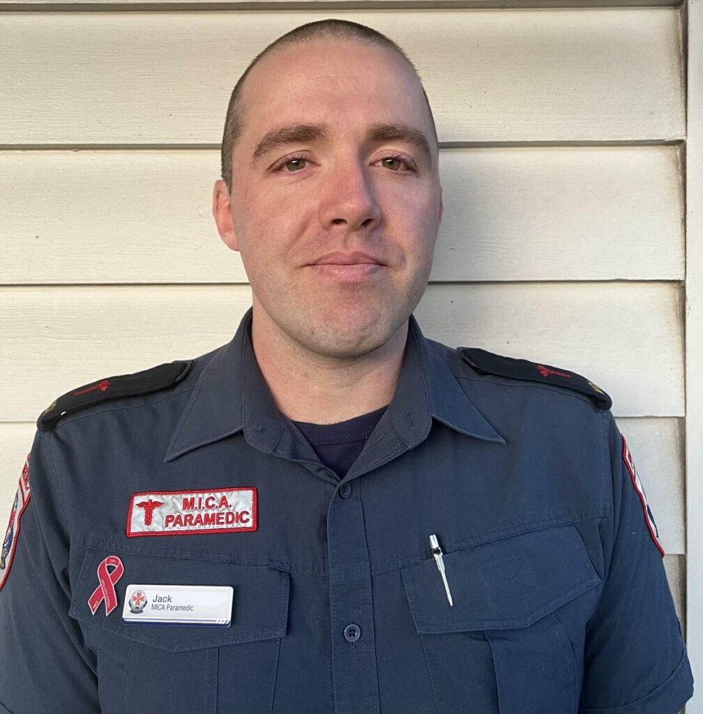
We discuss the phenomenon of CPR-induced consciousness (i.e. patients demonstrating awakeness during resuscitation) with Jack Howard, Intensive Care Paramedic at Ambulance Victoria in the northern suburbs of Melbourne, Australia, and first author on a recent literature review and Delphi-derived expert guideline on CPRIC management.
Takeaway lessons
- Data is light, but perhaps 1% of cardiac arrests have some form of consciousness witnessed.
- It is primarily a problem because of the potential to delay or interfere with care (either due to the emotional confrontation and surprise, or from actual physical interference with medical care). However, there are also ethical questions about patient suffering.
- The first response in many people seeing CPRIC will be to stop CPR and assume they’ve made a mistake about loss of pulses.
- CPRIC is associated with better outcomes, probably as a marker of better neurologic perfusion before and/or during arrest.
- There was general agreement by the panel that ketamine should be used as first-line for CPRIC. If unavailable or if it fails, the group was unable to agree on a best second line; fentanyl, midazolam, or a paralytic are all options. In CPRIC that physically interferes with care, larger doses are appropriate.
- Paralytics as a first line (without sedation) are never recommended.
- There is minimal data on the effect on outcomes when CPRIC is treated. One small Ambulance Victoria study had a trend towards lower rates of ROSC when sedation was used.
- Speak to patients as though they can hear and understand you.
- It is not clear but very possible that a larger number of patients than those who demonstrate external awareness may have a degree of subclinical consciousness; interviews of survivors and EEG analysis has supported this.
- Many CPRIC patients will have ROSC, but if they don’t, they are probably excellent candidates for ECPR/ECMO or other rescue interventions. A minimum of 45 minutes of resuscitation should be offered.

References
- The guideline: Howard J, Grusd E, Rice D, et al. Development of an international prehospital CPR-induced consciousness guideline: A Delphi study. Paramedicine. 2023;0(0). doi:10.1177/27536386231215608
- The preliminary scoping review: Howard J, Lipscombe C, Beovich B, Shepherd M, Grusd E, Nudell NG, Rice D, Olaussen A. Pre-hospital guidelines for CPR-Induced Consciousness (CPRIC): A scoping review. Resusc Plus. 2022 Nov 28;12:100335. doi: 10.1016/j.resplu.2022.100335. PMID: 36465817; PMCID: PMC9713363.
- The study mentioned about awareness during CPR: Parnia S, Keshavarz Shirazi T, Patel J, Tran L, Sinha N, O’Neill C, Roellke E, Mengotto A, Findlay S, McBrine M, Spiegel R, Tarpey T, Huppert E, Jaffe I, Gonzales AM, Xu J, Koopman E, Perkins GD, Vuylsteke A, Bloom BM, Jarman H, Nam Tong H, Chan L, Lyaker M, Thomas M, Velchev V, Cairns CB, Sharma R, Kulstad E, Scherer E, O’Keeffe T, Foroozesh M, Abe O, Ogedegbe C, Girgis A, Pradhan D, Deakin CD. AWAreness during REsuscitation – II: A multi-center study of consciousness and awareness in cardiac arrest. Resuscitation. 2023 Oct;191:109903. doi: 10.1016/j.resuscitation.2023.109903. Epub 2023 Jul 7. PMID: 37423492.