Episode 4: Venous congestion with Philippe Rola

Intensivist and passionate slayer of venous congestion Philippe Rola (@thinkingcc) shows us how to deresuscitate the septic patient, with guidance from his handy ultrasound.

Takeaway lessons

  1. Fluid overload is harmful and should be actively reduced, even in a patient in active shock; it will not harm them.
  2. The VEXUS exam is a good method for stratifying fluid overload by severity, i.e. severe (and harmful) versus mild (and relatively benign).
  3. The IVC, CVP, or hepatic vein doppler offer similar information, and are all effective means of assessing central venous pressure, the first and most important step in evaluating for venous congestion. The portal vein doppler offers the most additional diagnostic yield on top of this. Renal vascular doppler acts mostly as a “tiebreaker” when these other studies are equivocal.
  4. No one study or datapoint tells the whole story in these patients. Gather data from as many sources as possible to form the clearest picture.

Our apologies for the section of missing audio and slightly below-par audio quality in this one.

Resources

References

Beaubien-Souligny W, Benkreira A, Robillard P, et al. Alterations in Portal Vein Flow and Intrarenal Venous Flow Are Associated With Acute Kidney Injury After Cardiac Surgery: A Prospective Observational Cohort Study. J Am Heart Assoc. 2018;7(19):e009961.

Denault AY, Beaubien-Souligny W, Elmi-Sarabi M, et al. Clinical Significance of Portal Hypertension Diagnosed With Bedside Ultrasound After Cardiac Surgery. Anesth Analg. 2017;124(4):1109–1115.

Iida N, Seo Y, Sai S, et al. Clinical Implications of Intrarenal Hemodynamic Evaluation by Doppler Ultrasonography in Heart Failure. JACC Heart Fail. 2016;4(8):674–682.

Tang WH, Kitai T. Intrarenal Venous Flow: A Window Into the Congestive Kidney Failure Phenotype of Heart Failure? JACC Heart Fail. 2016;4(8):683–686. doi:10.1016/j.jchf.2016.05.009

Tremblay JA, Beaubien-Souligny W, Elmi-Sarabi M, et al. Point-of-Care Ultrasonography to Assess Portal Vein Pulsatility and the Effect of Inhaled Milrinone and Epoprostenol in Severe Right Ventricular Failure: A Report of 2 CasesA A Case Rep. 2017;9(8):219–223. [this pertains to a discussion of inhaled inodilators, which occurred during the lost section of audio]

Links

Credit and thanks to Eduardo R Argaiz (@ArgaizR) for this clip.

Episode 3: Refractory ARDS

What stops do you make along the garden path of hypoxic respiratory failure?

Resources

References

An ARDS review: Fan E, Brodie D, Slutsky A. Acute Respiratory Distress: Syndrome Advances in Diagnosis and Treatment. JAMA. 2018;319(7):698-710

Lung protective ventilation (The original ARDSnet ARMA trial): Acute Respiratory Distress Syndrome Network, Brower RG, Matthay MA, et al. Ventilation with lower tidal volumes as compared with traditional tidal volumes for acute lung injury and the acute respiratory distress syndrome. N Engl J Med. 2000;342(18):1301–1308.

Steroids for ARDS (SCCM/ESICM guidelines): Guidelines for the Diagnosis and Management of Critical Illness-Related Corticosteroid Insufficiency (CIRCI) in Critically Ill Patients (Part I): Society of Critical Care Medicine (SCCM) and European Society of Intensive Care Medicine (ESICM) 2017

Prone positioning (PROSEVA trial): Guérin C, Reignier J, Richard JC, et al. Prone positioning in severe acute respiratory distress syndromeN Engl J Med. 2013;368(23):2159–2168.

Driving pressure: Amato MB, Meade MO, Slutsky AS, et al. Driving pressure and survival in the acute respiratory distress syndromeN Engl J Med. 2015;372(8):747–755.

… and a driving pressure meta-analysis: Aoyama H, Pettenuzzo T, Aoyama K, Pinto R, et al. Association of Driving Pressure With Mortality Among Ventilated Patients With Acute Respiratory Distress Syndrome: A Systematic Review and Meta-Analysis. Crit Care Med. 2018;46(2):300–306.

APRV: Zhou Y, Jin X, Lv Y, et al. Early application of airway pressure release ventilation may reduce the duration of mechanical ventilation in acute respiratory distress syndromeIntensive Care Med. 2017;43(11):1648–1659.

Recruitment maneuvers (ART trial): Writing Group for the Alveolar Recruitment for Acute Respiratory Distress Syndrome Trial (ART) Investigators, Cavalcanti AB, Suzumura ÉA, et al. Effect of Lung Recruitment and Titrated Positive End-Expiratory Pressure (PEEP) vs Low PEEP on Mortality in Patients With Acute Respiratory Distress Syndrome: A Randomized Clinical Trial. JAMA. 2017;318(14):1335–1345.

Paralysis (PETAL trial): National Heart, Lung, and Blood Institute PETAL Clinical Trials Network, Moss M, Huang DT, et al. Early Neuromuscular Blockade in the Acute Respiratory Distress SyndromeN Engl J Med. 2019;380(21):1997–2008.

ECMO (EOLIA trial): Combes A, Hajage D, Capellier G, et al. Extracorporeal Membrane Oxygenation for Severe Acute Respiratory Distress SyndromeN Engl J Med. 2018;378(21):1965–1975.

Episode 2: Takotsubo (Stress) Cardiomyopathy

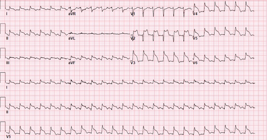
A tricky case of refractory heart failure.

Case files

Image credit: B Boling, personal files

Resources

References

Champion S, Belcour D, Vandroux D, et al. Stress (Tako-tsubo) cardiomyopathy in critically-ill patients. Eur Heart J. 2015;4(2):189-96.

Chockalingam A. Stress cardiomyopathy of the critically ill: Spectrum of secondary, global, probable and subclinical forms. Indian Heart J. 2018;70(1):177-84.

Dawson D. Acute stress-induced (takotsubo) cardiomyopathy. Heart. 2018;104:96-102.

Ghadri JR, Cammann VL, Jurisic S, Seifert B, Napp LC, Diekmann J, et al. A novel score (InterTAK Diagnostic Score) to differentiate takotsubo syndrome from acute coronary syndrome: results from the International Takotsubo Registry. Eur J Heart Fail. 2017;19:1036-42.

Muratsu A, Muroya T, Kuwagat Y. Takotsubo cardiomyopathy in the intensive care unit. Acute Med Surg. 2019;6(2):152-7.

Nunez-Gil IJ, Almendro-Delia M, Andres M, et al. Secondary forms of Takotsubo cardiomyopathy: A whole different prognosis. Euro Heart J. 2016;5(4):308-16

Episode 1: Necrotizing Soft Tissue Infection

A troubling case of a painful finger.

Resources

References

Links