Episode 28: Diuresis, deescalation, and liberation with Matt Siuba

How to take the well-resuscitated critically ill patient, get fluid out of them, deescalate their antibiotics, wean their sedation, reduce vent support, extubate, and get them out of the ICU—with Dr. Matt Siuba (Twitter: @msiuba), an intensivist at the Cleveland Clinic with an abiding interest in “zentensivism,” the art of doing less.

Takeaway lessons

  1. Portal vein pulsatility is a quick and useful addition to IVC assessment when evaluating volume status, particularly in ventilated patients where quick “eyeball” assessments of IVC variability is difficult (and measuring it is a pain). Consider pulse pressure variation too, in patients with a regular cardiac rhythm and on the vent.
  2. Start a journey of diuresis with furosemide, dose 40 times the serum creatinine. If you have a lot of work to do, or you expect resistance, add a thiazide (e.g. metolazone), and if more than a couple days of diuresis is expected, add spironoloctone as well to limit potassium wasting. Aim for >1–3 liters negative fluid balance per day, and generally schedule diuretics instead of manually spot dosing. Even if scheduled, however, follow up on urine output so you can increase the next dose if the last wasn’t adequate.
  3. Alternately if kidney function is quite poor, consider a furosemide stress test of 1 mg per kg of bodyweight (or 1.5 mg per kg for those with prior loop diuretic exposure). If output is poor, e.g. <200 ml in 2 hours, consider moving towards dialysis early.
  4. Tolerate a modest pressor increase induced by diuresis, such as norepinephrine within the 0.2 micrograms/kg range. However, volume overloaded patients usually tolerate volume removal fine, even if on pressors.
  5. Furosemide has a threshold and a ceiling, and there’s not much space between them, so feel free to give go fairly big in dosing without fear of complications.
  6. An elevated creatinine due to intrinsic renal injury is no contraindication to diuresis. In fact, it may improve with volume removal, if secondary to congestive nephropathy.
  7. Labs twice a day is fine for almost everyone, if labs are needed at all.
  8. Generally, don’t tap transudative pleural effusions solely for the purpose of fluid removal, except in non-intubated patients with very large effusions and respiratory distress.
  9. In general, wean FiO2 using pulse oximetry, not the blood gas.
  10. Even more than absolute finalization of cultures, deescalation of antibiotics should be informed by identification of the source and patient risk factors. A “surprise” resistant organism not predictable using these facts is a fairly uncommon event.
  11. Have a low threshold to switch stable, intubated patients to a pressure support mode. If fatiguing, consider giving more support instead of going to a control mode. PS of up to 8–15 cmH2O is very reasonable, and usually more comfortable (and less sedation-demanding) than forcing patients to accept a low tidal volumes on VCV. If a rate is needed, consider pressure control. A very variable tidal volume on PS may also be a good reason to give a set inspiratory time or volume.
  12. Night-time or late extubations are mostly dependent on staffing. If experienced providers are on hand, the clock should be no deterrent. If staffing is limited, it might give more pause. On the other hand, if your extubation practices are aggressive, most of these candidates were probably already extubated in the morning.
  13. Consider routinely extubating very obese patients to BiPap, and elderly patients (>65) or those with chronic cardiopulmonary diseases to HFNC. The latter is reasonable in most patients whose acute hypoxic disease has not yet fully resolved.
  14. When to downgrade patients from the ICU is less dependent on how long it’s been since extubation or getting off pressors, and more dependent on whether any disease process with the potential to worsen is still present (such as an uncontrolled source of sepsis).
  15. The most common cause of error in deescalation is doing it too slowly. Being “conservative” exposes patients to iatrogenic harm from prolonged intubation, antibiotics, lines, and ICU stays; that harm of omission isn’t better or safer than the alternate harms of commission. A patient who is admitted to the ICU, aggressively resuscitated, then rapidly improves and gets turned around isn’t moving “too fast”—that’s exactly the goal. Step up fast, step down fast.

Episode 27: Wilderness medicine with Ross Hofmeyr

Management of an alpine medicine scene including discussion of HAPE (High Altitude Pulmonary Edema), HACE (High Altitude Cerebral Edema), and suspension syndrome, with Dr. Ross Hofmeyr (@rosshofmeyr), anesthesiologist in the Department of Anaesthesia and Perioperative Medicine at the University of Cape Town, as well as cofounder and medical director of WildMedix, the oldest wilderness medicine support group in South Africa.

Takeaway lessons

  1. Scene safety comes first, which in a high-altitude setting includes consideration for the acclimatization of your team.
  2. Other than ABCs, think WMX:
    1. W= Factors of the Wilderness environment, such as altitude and exposure.
    2. M= Materials and Manpower; what’s needed for the rescue and what’s available?
    3. X= Exit strategy; from the start, think about how (and how rapidly) you’re getting out.
  3. Spinal stabilization is not feasible until a suspended patient can reach a stable location.
  4. Suspension syndrome (aka harness suspension syndrome, harness hang syndrome, suspension trauma) is a phenomenon of reduced venous return due to immobility while suspended in a vertical position (exacerbated by, although not requiring, a restrictive harness). Similar orthostasis can occur in healthy individuals forced to stand upright and immobile (at parade rest) without active muscle pumps, except they will then fall and self-reperfuse. In suspended patients, cardiac arrest can occur during suspension due to loss of preload, and further instability can occur after flow is restored due to a crush-syndrome-like reperfusion phenomenon with cold, acidotic blood. A hanging patient may be temporized by lifting their legs to a more horizontal position.
  5. Lowering a suspended patient is always easier than lifting them.
  6. If possible, consider deferring intubation in very austere settings, unless you realistically have the equipment, manpower, and space to transport a sedated, apneic patient while continuously ventilating them.
  7. Optic nerve sheath ultrasound can be a big diagnostic aid for ruling in cerebral edema in the setting of HACE or head trauma. A diameter >9–10 mm is suspicious for elevated ICP, >15 mm is a reliable rule-in, 6–9 mm is unclear. Differences between the two sides are concerning.
  8. The first treatment for any altitude sickness is oxygen; the second is descent. Everything else is temporizing or supportive.
  9. HACE is part of the spectrum of acute mountain sickness, and is caused by increased hydrostatic pressure and capillary permeability, leading to vasogenic edema. It tends to be caused by hypobaric hypoxemia, causing an afferent–efferent imbalance in cerebral vasculature.
  10. HAPE is caused by hypoxic pulmonary vasoconstriction and perhaps increased vascular permeability. It can cause severe hemorrhagic pulmonary edema.
  11. Give dexamethasone 8 mg IV (IM is okay too) for suspected HACE. No mannitol. Hypertonic saline can be considered but is poorly studied, and probably not smart while still far from help.
    1. Portable hyperbaric chambers can provide a substantial relative decrease in a patient’s altitude (the difference is more dramatic at higher altitudes), useful for temporization when immediate descent is impossible. However, they are bulky and completely envelope the patient, limiting further access for care. Provide other measures first, pressurize them therapeutically, then generally remove them from the bulky device for transport.
  12. Acetazolamide may help accelerate the acclimatization process, but can only do so much, and has no role in treating acute mountain sickness.
  13. >5,000 ft (1,500 m) or so is considered “high altitude” (some purists call 1,500–2,500 m/5,000–8,200 ft “intermediate altitude”). However, true altitude illness usually occurs above 3,500 m (11,500 ft), aka “very high altitude,” as above this level, it becomes impossible to hyperventilate enough to normalize alveolar oxygen concentration (PAO2).
  14. Above this level (~10,000 ft), gradual acclimatization is recommended. Sleeping altitude should only increase by about 500 m (1500–2000 ft) per day, and every second day should be a rest day.

Episode 26: ICU sedation, mobility, and delirium with Dale Needham

How to manage the intubated critically ill patient while keeping them awake, non-delirious, and mobile, with Dr. Dale Needham, FCPA, MD, PhD.

Dr. Needham is a Professor of Pulmonary and Critical Care Medicine as well as Physical Medicine and Rehabilitation at the Johns Hopkins University. He is also director of their Outcomes After Critical Illness and Surgery (OACIS) group, an attending intensivist in the medical intensive care unit, and the medical director of the Critical Care Physical Medicine and Rehabilitation program.

Takeaway lessons

  1. Start vent sedation with as-needed boluses of fentanyl alone. This is primarily for tube-related discomfort, with the assumption that patients may not need sedation per se unless they demonstrate the need. More fentanyl is often needed early on. If a long-acting paralytic was used for intubation, do consider a one-time benzo push or a larger opioid bolus for the initial period of paralysis.
  2. 25 mcg of fentanyl PRN every hour is a good starting point unless the patient is larger or opioid-tolerant. However, you can rapidly escalate the dose as needed. Even large bolus doses in a patient who has proven the need is preferable to routinely using a drip. Reserve the opioid drips for patients who require frequent boluses, and reserve sedatives for those who remain agitated after analgesia is in place.
  3. Soft restraints will often be used initially as patients emerge, but then may come off if patients prove to be well-oriented and cooperative. Very loose restraints are also an option over tightly restrictive restraints, serving only as a reminder.
  4. Mobility can start as soon as the day of intubation if the clinical picture and logistics are amenable.
  5. Well-trained nurses may be the best people to determine the need for additional sedation, restraints, sitters, etc. Restraints may sometimes worsen delirium and agitation; assess for their effects.
  6. Even in delirious patients, start with analgosedation (e.g. fentanyl)—these patients can’t tell you when they have pain, and pain is itself deliriogenic. After that, consider escalating to either dexmedetomidine, or PRN antipsychotics like haloperidol (which you can convert to oral quetiapine if it works).
  7. Remember that antipsychotics will neither prevent or treat delirium, and are only acting as a sedative for safety’s sake. If electing that approach, monitor QTc, start low (e.g. 1 mg haloperidol IV) and double it as needed until good effect is seen.
  8. Benzos are a last line, except in special circumstances (seizure, alcohol withdrawal) as they may worsen delirium, even if they temporarily hide it by extinguishing signs of agitation. Mostly, they defer the delirium to someone else’s shift. Hardly any patient goes from deep sedation to wakefulness without passing through a period of frank delirium.
  9. Mobilize early, even in patients modestly delirious and confused; it helps engage them, manages the pain of immobility, and tires them out to enable better sleep. It does require some attention towards safety by nursing and therapists; safety events related to mobility are minimal in good systems.
  10. It’s always hard to “escape” the vicious cycle of sedation and delirium once severe agitation is requiring multiple drips and deep sedation. Skilled nursing is key, with the shared understanding that sedation is harmful and needs to be (safely) weaned ASAP. Start with weaning the most deliriogenic medications, like benzos, then perhaps propofol. Expect a period of agitation and treat it with dexmedetomidine or PRN antipsychotics; use the approach of escalating antipsychotic as above, and if it works consider an oral antipsychotic for more steady state effect.
  11. “Chronic” ICU patients (stuck there for weeks or months, often due to disposition issues) can engage in highly aggressive PT/OT with multiple sessions for day. They are generally not delirious. SLP can attempt measures like in-line speaking valves or “talking trachs” and evaluate for swallowing while the trach is in place. Many such patients develop anxiety and cognitive issues, which is often best addressed via therapy (e.g. by rehab psychologists) rather than medications.
  12. In well-evolved systems with good practices around these areas, COVID-19 should ideally change things relatively little. Many patients can remain lightly sedated and mobile. Use video conferencing in lieu of visitation to redirect patients; you can even do it during a rehab session, using family as motivators and for redirection, and activity to keep them awake.
  13. Severe ARDS (e.g. from COVID) does take its toll. Proning can lead to shoulder injuries, and deep sedation and paralysis leaves severe physical and cognitive deficits. Agitation may lead to vent dyssynchrony leading to derecruitment. Try to have a slow, steady plan for weaning the vent and sedatives, and stick to it even across multiple shifts.

Resources

  1. 10th Annual Johns Hopkins Critical Care Rehabilitation Conference, November 4–6 2021, virtual format. Extensive material from prior conferences is available here as well.
  2. Staying Woke: review of sedation, mobility, and delirium
  3. Hopkins OACIS group
  4. Hopkins: Activity and Mobility Promotion
  5. Hopkins toolkit for early rehab programs

References

  1. Strom et al. “No sedation on the vent” trial
  2. Kollef et al. “Bolus vs continuous sedation” trial
  3. Hager et al. Reducing deep sedation and delirium in acute lung injury patients: a quality improvement project
  4. Needham et al. Early physical medicine and rehabilitation for patients with acute respiratory failure: a quality improvement project
  5. An example of nurse-controlled analgesia pumps

Episode 25: Rhabdomyolysis

Initial workup, fluid management, renal replacement, and other subtleties of caring for the critically ill patient with rhabdomyolysis.

Takeaway lessons

  1. Rhabdomyolysis is defined by elevated levels of creatinine kinase and/or myoglobin in the serum secondary to skeletal muscle breakdown with release of cellular contents. Common causes are crush or compartment syndrome, prolonged downtime on hard ground in patients who fell and cannot stand back up, and a variety of less common phenomena.
  2. Monitor with serum CK and/or myoglobin every 4–12 hours. Urine myoglobin is usually elevated, and AST/ALT, troponin, LDH, and potassium are all commonly high as well.
  3. The mainstay of treatment is vigorous hydration to flush the kidneys and prevent nephrotoxicity from myoglobin precipitation in the tubules. Historically this was via sodium bicarbonate (as myoglobin is less likely to precipitate in an alkaline environment) and forced diuresis with mannitol. These can be considered, but isotonic crystalloid may be as good in most cases.
  4. Do consider bicarb in the most severe cases, particularly if renal failure seems incipient. Maybe consider diuresis if the urine output is poor and fluid balance is increasingly positive. If oliguric due to AKI, consider reducing fluids, as the goal is urine output, not hypervolemia.
  5. Consider trending CK until it peaks, which may require dilution by the lab if it exceeds the upper limit of their assay. A CK that continues to rise may indicate an ongoing source of muscle injury. Consider trending urine pH as well if alkalinizing the urine with bicarb, targeting a pH >6.5.
  6. The lion’s share of rhabdo is mild or moderate, and often an incidental finding in the setting of other illness or injury. Occasional cases are severe and high risk for renal failure. The McMahon score can be used to try and predict these cases.
  7. In general, the role of hemodialysis or CVVH is the same as for anyone: renal replacement if kidney injury progresses to the point where it’s indicated. Some experimental work is underway with super high-flux CVVH filters which may help clear myoglobin, but in general CRRT has no disease-specific effects on rhabdo.

Special episode: Surviving COVID-19 with Eve Leckie


The COVID-19 pandemic will hopefully wind down this year. What happens next with these patients? A powerful discussion with Eve Leckie (@browofjustice), RN, CCRN, formerly of the CVCC at Dartmouth-Hitchcock and now disabled after contracting COVID. Learn about their acute course of illness, the challenges of navigating the healthcare system with this new disease, and their persistent, poorly-understood symptoms.

Takeaway lessons

  1. For Eve, orthostasis, heart rate and blood pressure abnormalities (e.g. POTS), and severe persistent fatigue, weakness, and “brain fog” are hallmarks of their chronic symptomatology. Shortness of breath, nerve-type symptoms (paresthesias, twitching), and intermittent fevers also continue to occur without a clear cause. Steroid courses seem to bring relief from some symptoms.
  2. Ground glass opacities and moderate airway thickening persists on imaging. In Eve’s case, their baseline asthma no longer responds to bronchodilators.
  3. Some “long haulers” have developed a criterion that most patients will have improvement in symptoms by 4 months post-infection, but there is a subset who does not fit that model.
  4. Patients who were advised to avoid healthcare, presume positivity, and defer testing now may face difficulty “proving” they had a definite infection for purposes like continued care or disability claims.
  5. Lack of understanding of this disease leads to a lack of diagnostic “boxes” to categorize symptoms, which often leads to dismissal and “non-diagnoses” of functional disorders or conversion syndrome. Until the scientific model is understood and diagnostic criteria have been validated, all that’s available is patient experiences, which should be trusted.
  6. Practical challenges like fatigue and cognitive issues can make dealing with healthcare, insurance, and supporting one’s self and family almost impossible without substantial assistance.

Links

Episode 24: Rural retrieval medicine with Minh Le Cong

Scene management, logistics, and stabilization of a blunt trauma patient in the Australian outback with Dr. Minh Le Cong (@ketaminh), rural GP and retrieval physician for the Royal Flying Doctor Service and host of the PHARM podcast.

Takeaway lessons

  1. If there is reasonable suspicion of the presence of a pneumothorax (of any size), have a low threshold to empirically decompress it before bringing the patient to altitude. Needles are okay, chest tubes are probably better.
  2. Hemothorax may be a soft reason to avoid chest decompression, which could conceivable remove the tamponading effect of intrapleural pressure.
  3. Consider ketamine for a neuro-stable induction.
  4. Abdominal aortic compression may be a salvage temporizing measure for penetrating abdominal/pelvic trauma where surgical intervention is delayed.
  5. Packaging depends on how much time you can afford to spend on scene. Simplify spinal precautions; consider measures like vacuum boards.
  6. All gasses expand at altitude and contract with descent. Think ETT cuffs, vacuum mattresses, pneumothoraces, etc, all which may shift and change size during travel.
  7. Whether practiced by paramedics, nurses, physicians, APPs, or others, transport and prehospital medicine is fundamentally the same business. The invasiveness of management may vary, but the core principles and the bulk of the approach does not. The addition of point-of-care ultrasound may be the biggest added value that would require higher levels of training.

Episode 23: COVID-19 pneumonia with Nicole King

Best practices in care of the critically ill patient with COVID-19 pneumonia are not known at this time. However, practical lessons from the ground are filtering in from those who have seen many of these patients, and Dr. Nicole King—critical care anesthesiologist, ECMO-ologist at the University of Cincinnati, and alumnus of the New York City COVID surge—is here to walk us through her experiences.

Takeaway lessons

  1. Do your swabs, cultures, and antigens to check for other viral and atypical pneumonias; other diseases still exist.
  2. Check a D-dimer to stratify hypercoagulability.
  3. Worsening tachypnea, distress, and ventilatory (not hypoxic) failure are a marker to upgrade care and/or intubate a borderline COVID patient.
  4. Treat initially with steroids per the RECOVERY trial (dexamethasone 6 mg daily for 10 days); then, if needing ICU or especially if needing intubation, consider the DEXA-ARDS protocol (20 mg daily for 5 days, then 10 mg daily for another 5 days). You may consider remdesevir or convalescent plasma at this point in the science, but don’t expect too much effect.
  5. Very prolonged courses on non-invasive positive pressure ventilation (CPAP/BiPAP) may be more acceptable here than in other diseases; although not very appealing it may be preferable to intubation. Try cycling on and off to improve tolerability. Consider an NG tube for nutrition.
  6. However, beware of profoundly large tidal volumes in spontaneously breathing patients due to their remarkable air hunger, which may predispose to lung injury—spontaneous pneumothorax can occur even in these non-invasive patients.
  7. When air hunger is profound, you may need to decide whether to sedate them to control tidal volumes, or allow (potentially harmful) large volumes, or try to limit them with the vent with the possible result of a dyssynchronous and air hungry patient.
  8. It’s not clear whether intubation worsens outcomes, but it’s clear that the patients who require intubation seem to do very poorly; part of the reason may be selection for high-risk patients as we try to avoid intubating when possible, but part may be iatrogenesis from things like sedation and paralysis.
  9. PEEP is not very high-yield on many of these patients—either they need little recruitment (good compliance) or their compliance is so poor they are minimally recruitable—but since they are frequently so borderline, they still often end up on high PEEPs because they need whatever little margin of recruitment it does provide.
  10. APRV makes sense and may have a role, but you will generally need to avoid paralysis and lighten sedation, as CO2 can become very hard to manage without significant spontaneous breathing.
  11. Prone early, when the lungs are still recruitable and salvageable. It’s unclear whether it’s beneficial for lung protection even when the benefit on oxygenation is not impressive. It can be logistically challenging due to obesity and hemodynamic instability.
  12. If considering ECMO, do it early. A prolonged course (intubated >10–14 days or on high vent settings for >7 days) is a contraindication, as permanent lung injury has already set in and recovery is less likely. To achieve this, aggressively manage early with usual methods, then if they seem to be refractory, consider ECMO as soon as the trajectory is clear—don’t give them a “waiting period” of days/weeks of vent failure first.
  13. VV ECMO is usually adequate, particularly if you cannulate early enough that cardiovascular collapse has not set in, and may limit the iatrogenic harms. Whether VA ECMO may have a role for those with PE and RV strain is unclear.
  14. Early tracheostomy is reasonable, but it’s not a panacea—most patients still end up needing significant vent support, sedation, etc.
  15. 20 days after their initial symptom onset, patients are likely no longer infectious and isolation precautions can be withdrawn, regardless of testing.
  16. During and perhaps for a prolonged period after their peak illness, some of these patients will have a persistently elevated respiratory drive—tachypnea, high minute ventilations and work of breathing—that may not portend anything acutely untoward, or at least may not be preventable.
  17. When patients start looking worse, look at the right heart, which will often be strained from high PEEP and/or the presence of PEs. Treat the latter, and consider inhaled pulmonary vasodilators.

Episode 22: Aneurysmal subarachnoid hemorrhage with Thomas Lawson

Initial approach, supportive care, risk stratification, and management of the troublesome complications for ruptured subarachnoid aneurysm, with Thomas Lawson (@TomLawsonNP), nurse practitioner in the neurocritical care unit at OSU Wexner Medical Center.

Takeaway lessons

  1. SAH + shock or hypoxemia = suspect neurogenic pulmonary edema and/or Takotsubo cardiomyopathy.
  2. Aneurysmal SAH is much different from traumatic and other etiologies of SAH. Not only does it require securing the aneurysm (early rebleeding is associated with substantially increased mortality), it conveys generally greater morbidity.
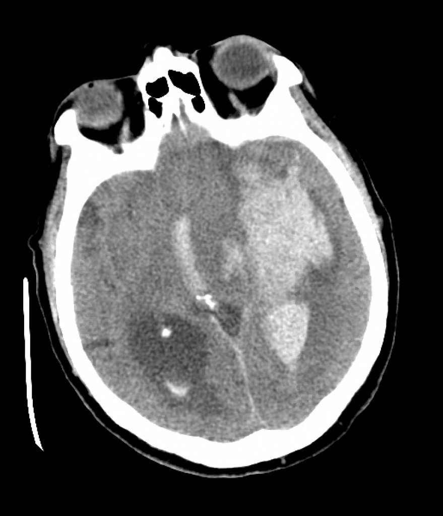
  3. The “hanging chicken sign” at the base of the skull (across the basilar cisterns) is the most distinctive SAH finding on head CT. Look also for associated IPH or IVH, as well as signs of hydrocephalus, such as third ventricle enlargement or presence of a temporal horn in the lateral ventricles (normally just a faint C-shaped slit).
  4. Use either the Hunt and Hess or WFNS scores to prognosticate mortality, while considering the Modified Fischer score to assess risk of vasospasm, a complication usually occurring between 3–14 days (sometimes up to 21) days after onset of symptoms.
  5. With any acute neuro changes in the SAH patient, strongly consider the “stat stat” head CT to assess for rebleeding or worsening hydrocephalus. Also assess for functioning of an EVD, as an obstructed tube may indicate hydrocephalus as well.
  6. For deterioration in later timeframes, go with a CTA (or transcranial dopplers) to assess for vasospasm. If present, talk to your neurointerventionalists about offering catheter-directed vasodilators, such as verapamil or nicardipine; also, augment the BP to perfuse past the spasm.
  7. Many patients who spasm will continue to spasm. Watch diligently as some may need repeated neuro-intervention. IV milrinone and intra-thecal nicardipine (into an EVD) are available as last-ditch efforts.
  8. CTA will not show thrombosed aneurysms; such patients need a catheter angiogram and/or repeat imaging.
  9. “Triple H therapy” for SAH—hypertension, hypervolemia, and hypernatremia (or hemodilution) is dead. Just shoot for reasonable hypertension, plus euvolemia. Target a normal MAP and SBP <220.
  10. Polyuria should prompt concern for cerebral salt wasting, which is often followed by vasospasm. It can be distinguished from SIADH by the presence of hypovolemia.
  11. If a patient has no seizures noted, a 3–7 day course of anti-epileptics (e.g. levetiracetam) is reasonable. If they do seize, continue for longer.
  12. EVDs can come out when the daily drainage is dwindling. Then wean by raising the pop-off by about 5 cm H2o per day, monitoring the neuro exam and perhaps CT scans. Once at 20 cm H2O, clamp it, then consider removal. Occasionally patients may have permanent dysfunction of their arachnoid granulations and need a VP shunt, although this must usually be placed in a delayed fashion to avoid blockage by proteinaceous CSF early on.

References

Consensus guidelines: Grasso G, Alafaci C, Macdonald RL. Management of aneurysmal subarachnoid hemorrhage: State of the art and future perspectives. Surg Neurol Int. 2017 Jan 19;8:11. doi: 10.4103/2152-7806.198738. Erratum in: Surg Neurol Int. 2017 Apr 26;8:71. PMID: 28217390; PMCID: PMC5288992.

IV milrinone: Lannes M, Teitelbaum J, del Pilar Cortés M, Cardoso M, Angle M. Milrinone and homeostasis to treat cerebral vasospasm associated with subarachnoid hemorrhage: the Montreal Neurological Hospital protocol. Neurocrit Care. 2012 Jun;16(3):354-62. doi: 10.1007/s12028-012-9701-5. PMID: 22528278.

Episode 21: Trauma resuscitation with Scott Weingart

A patient with multiple abdominal gunshot wounds, resuscitated before and after damage control surgery by the legendary Scott Weingart (@emcrit): emergency physician, surgical intensivist by way of Shock Trauma in Baltimore, director of an emergency critical care program, and longtime innovator in medical education and FOAM via the EMCrit podcast and blog.

Our 1st anniversary episode!

Takeaway lessons

  1. In an ideal world, penetrating abdominal trauma in an unstable patient would proceed directly to the OR with no delay by the ED. Resuscitation, obtaining access, and so forth can occur in the OR perioperatively.
  2. Have a low threshold for activating the massive transfusion protocol. (A positive FAST would reinforce that decision.)
  3. Venous access: an introducer sheath (Cordis) or dialysis catheter in the femoral or subclavian veins. Blind subclavian lines may be ideal but are a dying skill (ultrasound-guided subclavians are too slow). Femoral lines are somewhat less desirable with abdominal trauma as the IVC may be violated. Always use ultrasound for the fem. Ultrasound-guided IJ is okay if that’s all your people are comfortable with. Peripherals are okay, a RICC is better, but central access is most reliable.
  4. Arterial access: consider an 18g femoral arterial line in the common femoral artery, allowing BP monitoring but also wire insertion in case endovascular procedures are needed, such as REBOA or IR embolization. A 16g arterial is a bit over-large but okay. 20g is okay but needs upsizing later.
  5. Give TXA if within 1 hour (not 3 hours) of injury: 1 gram with the first unit of blood. (2 grams is an option too.) The need for the subsequent maintenance drip can be determined by TEG later.
  6. Not too much role for TEG in the initial ED presentation. Takes too long for the “clot stability” portion to develop anyway. But it’s useful after surgery, during the post-acute phase.
  7. A secondary CT scan, even performed after initial damage control surgery, can be very useful to localize non-obvious lesions which may require a secondary repair. If not performed, a careful clinical tertiary survey is mandatory to evaluate for extremity perfusion (i.e. a missed vascular injury) and other injuries not noted during surgical exploration.
  8. Labs are slow. Point of care labs are a little better. During the most acute period, mostly transfuse to target vitals (MAP >65 or so). As they begin to achieve hemostasis, things slow down, and it start to become possible to follow labs. Continue to use a balanced ratio unless you can use TEG to guide FFP and platelets.
  9. Check body temperature and use forced-air rewarming if needed.
  10. Some degree of distributive shock can result from the post-traumatic inflammatory (SIRS) response, especially downstream of the first hour or two. Vasopressin may be the first line agent for this, as many trauma patients are vasopressin-depleted. Still, assume all shock is hemorrhagic until proven otherwise. Skin temperature helps some, as vasodilation = warm but hypovolemia = cold. As volume is replaced, vasopressors come off, and perfusion overall improves; a “good” BP with ice-cold extremities is still in shock and still organ-injurious.
  11. Most importantly, shock is a vicious cycle, so the sooner you can correct hypoperfusion, the less secondary distributive shock and organ failure you’re likely to see down the road.
  12. Push calcium chloride to maintain a normal ionized calcium, which may have a nice BP effect as well.
  13. Use ultrasound (as windows allow), particularly to rule out the need for inotropes.
  14. Empiric cryo not a bad idea early. Later, use either TEG or labs, in which case target a fibrinogen level of >150 or even >200.
  15. Consider PCCs (KCentra) if: 1. FFP is delayed or unavailable; 2. Very behind in resuscitation (i.e. unbalanced resuscitation or lots of crystalloid so far); 3. Patient is on warfarin; 4. Cirrhotics.
  16. Activated Factor VII (NovoSeven) probably has no role at this point. It makes transient clots, but not durable ones, and causes pathologic thrombi; outcomes are poor. PCCs including activated VII is probably better, if you must.
  17. Autotransfusion (i.e. from chest tube drainage) is a logistical pain, clotty, and really only a substitute for PRBCs, since after hitting the container it contains almost no clotting factors or platelets. Usually not worth the bother. Banked whole blood would be much better, but is also a logistical challenge from a systems perspective.
  18. Serious ongoing bleeding in excess what can be matched with a properly-executed MTP is probably a soft indication to surgically reexplore, or at least consider imaging for missed injuries, since purely “medical” bleeding will infrequently be so brisk. Particularly when the initial exploration was in the setting of serious multi-trauma, missing an injury is common, particularly when they’re under-resuscitated (and hence “permissively” hypotensive) at the time of exploration.
  19. If you must give crystalloid instead of blood, consider hypertonic saline, preferably buffered.
  20. Tolerating a lower MAP (maybe down to 50–55) may be better than giving high doses of vasopressors or crystalloid while waiting for blood. If bleeding seems to have stopped, however, you might start treating hypotension as purely distributive shock and allow higher vasopressor doses.
  21. In a young trauma patient with good baseline condition, aggressive curative care is probably ethical almost ad infinitum. It’s often reasonable to continue efforts until they arrest and die or resources are exhausted.
  22. The truly sick trauma patient does not “take a joke,” and absolutely requires your undivided attention and the most astute, timely, on-the-ball care you can provide. Your normal “max effort” is not enough; like a lazy runner, you need to push and find your true limits in order to save these people.

References

  1. The ICU trauma resuscitation: More reading on the post-op resuscitation process.

Episode 20: Post-CABG emergencies with Kris Ramilo and Brendan Riordan

Looking at the workflow of a fresh post-op open heart surgery patient, as well as what to do when it devolves into cardiac tamponade, with (returning) guest Brendan Riordan, cardiothoracic ICU PA (@concernecus) at the University of Washington, and his NP colleague Kris Ramilo (@krsrml0).

[Audio quality was a bit dodgy in this one; sorry all!–eds.]

Takeaway lessons

  1. Handoff from the OR to ICU team is a tricky time, and should ideally be somewhat formalized, involve both surgical and anesthesia teams at the bedside, and including talk about what happened, what they expect, and things to look out for.
  2. The ERAS (Early Recovery After Surgery) protocol has entered the CT surgery world as ERACS, including post-op measures for typical cases such as reversal of neuromuscular blockade and a 4-hour target for extubation (versus the usual 6 hours). Most routine cases will aim to be “bloodless,” i.e. only transfusing autologous (“Cell Saver” blood), as transfusion of banked blood is considered an STS measure.
  3. Milking or stripping chest tubes during the immediate post-op period to maintain patency may or may not be necessary as a routine practice, but should certainly be attempted if output drops or tamponade is suspected.
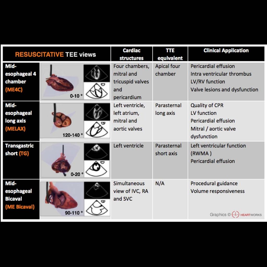
  4. Point-of-care TTE is always difficult in these patients, due to their windows being obscured by dressings; the apical 4-chamber will often be the most useful view. When seriously considering graft failure, TEE may be valuable to diagnose graft failure (by noting regional wall motion abnormality), as well as appreciating cardiac tamponade.
  5. As in all cases, tamponade in this setting is diagnosed by echocardiographic pericardial effusion plus signs of tamponade physiology (chamber collapse, etc), with the caveat that effusions may be loculated or unusual-appearing due to the recent violation of the pericardium.
  6. With a PA catheter, remember that narrowing of the PA pulse pressure and convergence of the CVP and PA pressures (particularly via a falling PA pressure) is classic for tamponade physiology.
  7. Resternotomy is not something to be undertaken lightly, but still must be performed immediately and aggressively when indicated. Close involvement with the surgical team is usually essential to make this decision.
  8. Adding an inotrope can be somewhat diagnostic, as patients with mere myocardial stunning will improve, whereas patients with tamponade will generally have little response.
  9. Give calcium chloride freely to the unstable post-cardiac surgery patient.
  10. Be aggressive with fluid resuscitation and consider epinephrine up to 0.06–0.08 mcg/kg/min or dopamine (if you must) up to 3–5 mcg/kg/minute.
  11. The purpose of reopening the chest is to avoid external chest compressions, which tend to abuse RCA grafts (which sit just below the sternotomy wires), and are generally ineffective anyway compared to internal cardiac massage, since the pressure chamber of the chest is not intact.
  12. The role of the ICU team while the surgical team reopens the chest is typically to assist with sedation, ventilator management, and hemodynamics. Anesthesia may or may not be present but is often performing TEE.
  13. Patients reopened at the bedside will often be left with open chests, and planned to return back to the OR for washout and closure the next day. Plan on prophylactic antibiotics (e.g. vancomycin).