Episode 9: Right heart failure and the SAVIOR protocol with Habib Srour (part 1)

The book

Buy the new textbook (Bryan edited, Brandon authored a chapter) here or on Amazon:

Concepts in Surgical Critical Care, First Edition

ed. Bryan Boling, DNP, ACNP; Kevin Hatton, MD, FCCM; Tonja Hartjes, DNP, ACNP-BC, CCRN, FAANP

The podcast

An in-depth look at the management of right heart failure, with a focus on preserving peri-intubation hemodynamics using the SAVIOR protocol—featuring its co-creator, anesthesiologist and intensivist from the University of Kentucky, Habib Srour.

Takeaway lessons

  1. When facing undifferentiated shock and a complex picture, look for one point of data to help distinguish the etiology. Try touching the feet: cold is a good indicator of a significant cardiogenic component.
  2. The flip side of hypoxic vasoconstriction is hyperoxic vasodilation of the pulmonary vasculature—i.e. an overly high FiO2 will tend to worsen V/Q matching.
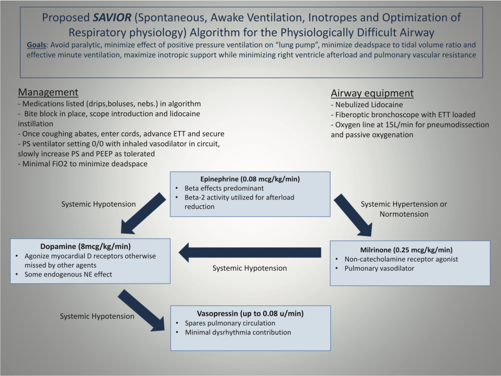
  3. To hemodynamically manage RV failure without worsening RV afterload, consider the Rule of 8s cocktail:
    1. Epinephrine .08 mcg/kg/min
    2. Dopamine 8 mcg/kg/min
    3. Vasopressin .08 units/min
    4. Inhaled epoprostenol (Veletri/Flolan) 8 ml/hr
  4. The “lung pump” of negative pressure respiration provides a substantial amount of cardiac output, particularly in the setting of RV failure. Paralysis, sedation, and intubation removes this. The period of apnea also worsens acidosis which increases PVR.
  5. The dead space to tidal volume ratio increases by at least 50% after intubation; it will be impossible to match an already-high spontaneous minute ventilation on the ventilator.

Resources

The SAVIOR algorithm. Figure 1 from Srour et al (vide infra).

References

Episode 8: Palliative care with Jessica McFarlin (part 2)

The second part of our look at a case of catastrophic intracranial hemorrhage, with a focus on goals of care, family interaction, prognostication, and other end-of-life aspects, with neurointensivist and palliative care physician Jess McFarlin (@JessMcFarlinMD).

See Part 1 here.

Takeaway lessons

  1. Useful phrase: “Can I tell you what to expect during the dying time?”
    1. Discuss the possibility of secretions, etc. Use glycopyrrolate.
  2. Use opioids if you expect dyspnea, otherwise not always needed. Can try a pressure support trial on the ventilator to get a sense for tachypnea.
  3. Let both family and the nurse know what to expect after extubation.
  4. Other than the occasional incidence of troubling myoclonus with fentanyl, and restrictions on its use outside of the ICU in many centers, all opioids are probably equally good for end-of-life care. Consider hydromorphone in renal patients.
  5. In general, stop tube feeds at the end of life, and stop trying to ensure full nutrition, but do offer food and drink for comfort. Dying tends to limit hunger and caloric needs anyhow. Stop IV fluids as well.
  6. When families invoke a “miracle scenario,” reframe by asking what a miracle might look like for them, or raise the possibility that the miracle won’t be survival, but another outcome such as surviving until the rest of the family arrives, or being comfortable and pain-free during the dying process.
    1. Use “I wish [it would work]” statements to express empathy and a shared perspective, while maintaining a fact-based reality. Stop there and don’t wade into details.
    2. Turn miracles into concrete plans by establishing a time trial with a deadline, with clear markers for what success will look like.

Episode 7: Palliative care with Jessica McFarlin (part 1)

Neurointensivist and palliative care physician Jess McFarlin (@JessMcFarlinMD) walks us through a case of catastrophic intracranial hemorrhage, with a focus on goals of care, family interaction, prognostication, and other end-of-life aspects.

See Part 2 here.

Takeaway lessons

  1. We can undo most things except death, so in most cases, a short trial (perhaps 3 days) of fully aggressive care after an ICU admission is reasonable to help clarify the eventual prognosis. Set clear guideposts for when you’ll regroup to make more decisions about the direction of care.
  2. The ICH score is a helpful guide for early prognostication in spontaneous intraparenchymal hemorrhage.
  3. When prognosticating, express the range of outcomes in terms of three possibilities, as determined by the currently available data: the best case, the worst case, and the most likely case.
  4. Useful questions for families:
    1. If your [loved one] could hear this prognosis, what would he/she say is most important to him/her?
    2. What’s most important to you at this stage? What are you most afraid of?(Many are more concerned about discomfort, pain, or “struggling” at the end of life than about the prospect of death itself.)
  5. When you’re asked, “What would you do if it were your mom?” the question is not really about your mom; it’s a request for a recommendation about theirs.
  6. Transition from open-ended questioning, and translate their values/goals into an actionable plan, by using alignment statements: “What I’m hearing you say/it sounds like…” leads to “Hearing that, may I make a recommendation?”

Resources

References

  • ICH score: mortality prediction score for spontaneous intraparenchymal hemorrhage
  • Vital Talk: training resources for the skills of executing palliative and end-of-life conversations

[this episode was reposted on the website 4/27/2020 due to a database reversion after an unfortunate system breach —ed.]

Episode 6: Status epilepticus with Gracia Mui

Neurologist and neurointensivist Gracia Mui shows us the workup, initial management, and escalation of care for a case of refractory status epilepticus.

Takeaway lessons

  1. First-time unprovoked seizures usually need no further workup except screening for an underlying trigger, such as a tox screen, basic chemistries, and imaging as appropriate.
  2. Initial seizure therapy: wait around 5 minutes, then give 2–4 mg lorazepam. Repeat every minute or so until convulsions stop, up to 0.1 mg/kg total.
  3. Give an anti-epileptic concurrently: fosphenytoin or phenytoin (15–20 mg/kg), levetiracetam, or valproic acid (40 mg/kg, up to 3000 mg) are all acceptable. If using levetiracetam (Keppra), give a real dose of 60 mg/kg (max of 4.5 g).
  4. After loading with benzos and/or anti-epileptics, if convulsions stop and the patient remains unresponsive, consider the duration of the drug you used. If it’s wearing off (e.g. after about an hour for lorazepam) and they remain unresponsive, suspect non-convulsive status epilepticus.
  5. Any patient not waking up needs an EEG. If not available, they may need empiric deep sedation and intubation until EEG can prove the absence of seizures.
  6. If convulsions are absent, that’s good, as convulsive seizures are more harmful than non-convulsive, but not as good as obviating seizure activity on EEG.
  7. Other than the practical, there is no upper limit for benzodiazepine dosing.
  8. Once you’ve successfully achieved the desired EEG result (either burst suppression or simply the absence of seizure activity) using anesthetics, hold them for about a day, then lighten sedation to see if seizures recur. If so, re-deepen sedation (perhaps for twice as long), increase anti-epileptic agents, then try again.
  9. The patient in status should routinely be screened for underlying triggers, including brain imaging and LP (remember autoimmune causes such as NMDA encephalitis). But about half the time, even in severe refractory status, no underlying cause will be identified.

Resources

References

[this episode was reposted on the website 4/27/2020 due to a database reversion after an unfortunate system breach —ed.]

Episode 5: Cardiogenic shock and ECMO with Brendan Riordan

Cardiothoracic critical care PA Brendan Riordan (@concernecus) shows us his initial approach to the patient in cardiogenic shock, including initiating mechanical support, managing ECMO (plus Impella), and eventual weaning and discontinuation of support.

Some pearls

  1. Anticoagulation on VA ECMO can be titrated to bleeding risk, with a balance between bleeding and circuit longevity—the latter being more than an inconvenience, as changing the circuit in a patient fully dependent on the pump is fraught. Anti-Xa levels are more reliable than the PTT. In a patient with HIT, you may be able to treat through it with bivalirudin, as the heparin-bonded circuit usually cannot be switched out.
  2. “Hypoxemia” on VA ECMO is either regional hypoxemia/North-South syndrome/harlequin syndrome, or oxygenator failure. Rule out the latter by checking a post-oxygenator ABG or just looking to ensure the outflow blood is bright red. Rule in the former by evaluating the ABG or SpO2 from the right upper extremity.
  3. Preemptively placing an anterograde perfusion catheter in the femoral artery is not absolutely mandatory, but is probably simpler and perhaps safer than placing one reactively.
  4. A PA catheter is more useful for weaning ECMO than during the period of full support.
  5. Readiness for weaning is evaluated by recognition of improving cardiac pulsatility, followed by a trial of weaning down pump flow, and finally decannulation in the OR. Consider leaving the Impella if there are any lingering concerns.

Resources

References

Special episode: Initial vent settings for COVID-19

A 5-minute episode describing three hyper-simple, generally safe recipes for the initial strategy of mechanical ventilation after intubating a COVID-19 patient.

Caveat: this is intended for trained clinicians, such as emergency medicine providers, who already have a general understanding of safe and sound life support practices. It glosses over a great deal and is not meant as a primer for trainees.

Resources

Full ARDSnet protocol

Special episode: COVID-19 brief

A fast-tracked update: what we know about the active pandemic of the novel coronavirus (SARS-CoV-2) and its resulting respiratory syndrome, COVID-19.

Takeaway lessons

  • Treat like viral pneumonia/ARDS. High PEEP. Prone early. Keep fluid balance dry.
  • Probably avoid CPAP/BiPAP except perhaps for very short trials. Unclear role for HFNC.
  • Watch out for myocarditis-type picture with cardiogenic shock and arrhythmias.
  • Take isolation extremely seriously.
  • Think about innovative ways to optimize patient flow.
  • Take care of yourself and each other, and try to stay sane.

Resources

References

Episode 4: Venous congestion with Philippe Rola

Intensivist and passionate slayer of venous congestion Philippe Rola (@thinkingcc) shows us how to deresuscitate the septic patient, with guidance from his handy ultrasound.

Takeaway lessons

  1. Fluid overload is harmful and should be actively reduced, even in a patient in active shock; it will not harm them.
  2. The VEXUS exam is a good method for stratifying fluid overload by severity, i.e. severe (and harmful) versus mild (and relatively benign).
  3. The IVC, CVP, or hepatic vein doppler offer similar information, and are all effective means of assessing central venous pressure, the first and most important step in evaluating for venous congestion. The portal vein doppler offers the most additional diagnostic yield on top of this. Renal vascular doppler acts mostly as a “tiebreaker” when these other studies are equivocal.
  4. No one study or datapoint tells the whole story in these patients. Gather data from as many sources as possible to form the clearest picture.

Our apologies for the section of missing audio and slightly below-par audio quality in this one.

Resources

References

Beaubien-Souligny W, Benkreira A, Robillard P, et al. Alterations in Portal Vein Flow and Intrarenal Venous Flow Are Associated With Acute Kidney Injury After Cardiac Surgery: A Prospective Observational Cohort Study. J Am Heart Assoc. 2018;7(19):e009961.

Denault AY, Beaubien-Souligny W, Elmi-Sarabi M, et al. Clinical Significance of Portal Hypertension Diagnosed With Bedside Ultrasound After Cardiac Surgery. Anesth Analg. 2017;124(4):1109–1115.

Iida N, Seo Y, Sai S, et al. Clinical Implications of Intrarenal Hemodynamic Evaluation by Doppler Ultrasonography in Heart Failure. JACC Heart Fail. 2016;4(8):674–682.

Tang WH, Kitai T. Intrarenal Venous Flow: A Window Into the Congestive Kidney Failure Phenotype of Heart Failure? JACC Heart Fail. 2016;4(8):683–686. doi:10.1016/j.jchf.2016.05.009

Tremblay JA, Beaubien-Souligny W, Elmi-Sarabi M, et al. Point-of-Care Ultrasonography to Assess Portal Vein Pulsatility and the Effect of Inhaled Milrinone and Epoprostenol in Severe Right Ventricular Failure: A Report of 2 CasesA A Case Rep. 2017;9(8):219–223. [this pertains to a discussion of inhaled inodilators, which occurred during the lost section of audio]

Links

Credit and thanks to Eduardo R Argaiz (@ArgaizR) for this clip.

Episode 3: Refractory ARDS

What stops do you make along the garden path of hypoxic respiratory failure?

Resources

References

An ARDS review: Fan E, Brodie D, Slutsky A. Acute Respiratory Distress: Syndrome Advances in Diagnosis and Treatment. JAMA. 2018;319(7):698-710

Lung protective ventilation (The original ARDSnet ARMA trial): Acute Respiratory Distress Syndrome Network, Brower RG, Matthay MA, et al. Ventilation with lower tidal volumes as compared with traditional tidal volumes for acute lung injury and the acute respiratory distress syndrome. N Engl J Med. 2000;342(18):1301–1308.

Steroids for ARDS (SCCM/ESICM guidelines): Guidelines for the Diagnosis and Management of Critical Illness-Related Corticosteroid Insufficiency (CIRCI) in Critically Ill Patients (Part I): Society of Critical Care Medicine (SCCM) and European Society of Intensive Care Medicine (ESICM) 2017

Prone positioning (PROSEVA trial): Guérin C, Reignier J, Richard JC, et al. Prone positioning in severe acute respiratory distress syndromeN Engl J Med. 2013;368(23):2159–2168.

Driving pressure: Amato MB, Meade MO, Slutsky AS, et al. Driving pressure and survival in the acute respiratory distress syndromeN Engl J Med. 2015;372(8):747–755.

… and a driving pressure meta-analysis: Aoyama H, Pettenuzzo T, Aoyama K, Pinto R, et al. Association of Driving Pressure With Mortality Among Ventilated Patients With Acute Respiratory Distress Syndrome: A Systematic Review and Meta-Analysis. Crit Care Med. 2018;46(2):300–306.

APRV: Zhou Y, Jin X, Lv Y, et al. Early application of airway pressure release ventilation may reduce the duration of mechanical ventilation in acute respiratory distress syndromeIntensive Care Med. 2017;43(11):1648–1659.

Recruitment maneuvers (ART trial): Writing Group for the Alveolar Recruitment for Acute Respiratory Distress Syndrome Trial (ART) Investigators, Cavalcanti AB, Suzumura ÉA, et al. Effect of Lung Recruitment and Titrated Positive End-Expiratory Pressure (PEEP) vs Low PEEP on Mortality in Patients With Acute Respiratory Distress Syndrome: A Randomized Clinical Trial. JAMA. 2017;318(14):1335–1345.

Paralysis (PETAL trial): National Heart, Lung, and Blood Institute PETAL Clinical Trials Network, Moss M, Huang DT, et al. Early Neuromuscular Blockade in the Acute Respiratory Distress SyndromeN Engl J Med. 2019;380(21):1997–2008.

ECMO (EOLIA trial): Combes A, Hajage D, Capellier G, et al. Extracorporeal Membrane Oxygenation for Severe Acute Respiratory Distress SyndromeN Engl J Med. 2018;378(21):1965–1975.

Episode 2: Takotsubo (Stress) Cardiomyopathy

A tricky case of refractory heart failure.

Case files

Image credit: B Boling, personal files

Resources

References

Champion S, Belcour D, Vandroux D, et al. Stress (Tako-tsubo) cardiomyopathy in critically-ill patients. Eur Heart J. 2015;4(2):189-96.

Chockalingam A. Stress cardiomyopathy of the critically ill: Spectrum of secondary, global, probable and subclinical forms. Indian Heart J. 2018;70(1):177-84.

Dawson D. Acute stress-induced (takotsubo) cardiomyopathy. Heart. 2018;104:96-102.

Ghadri JR, Cammann VL, Jurisic S, Seifert B, Napp LC, Diekmann J, et al. A novel score (InterTAK Diagnostic Score) to differentiate takotsubo syndrome from acute coronary syndrome: results from the International Takotsubo Registry. Eur J Heart Fail. 2017;19:1036-42.

Muratsu A, Muroya T, Kuwagat Y. Takotsubo cardiomyopathy in the intensive care unit. Acute Med Surg. 2019;6(2):152-7.

Nunez-Gil IJ, Almendro-Delia M, Andres M, et al. Secondary forms of Takotsubo cardiomyopathy: A whole different prognosis. Euro Heart J. 2016;5(4):308-16